
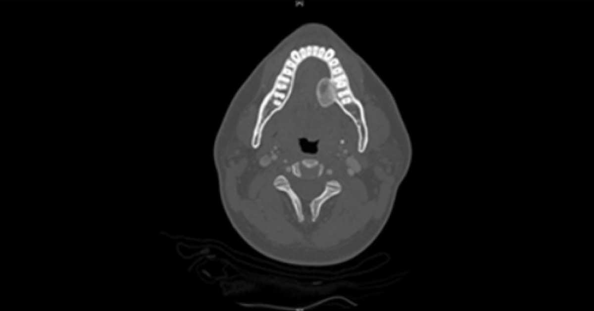
A 28-year-old male presented with an exophytic lingual plate expansion in the left posterior mandible (image 1). Image description: 1: A panoramic radiograph taken at the first presentation, demonstrating a large radiopaque lesion in the left posterior mandible projecting as a well demarcated radiopaque lesion. History of present illness The panoramic radiograph demonstrates a well-defined exophytic radioplaque lesion protruding into the floor of mouth and measuring approximately 4×2 cm. The lesion had grown gradually since the patient was 13-16 years of age, and has not been accompanied by pain or bleeding.
The associated teeth are vital. The lesion was surgically removed, and recurred within a year (image 2). Image description: 2: A radiographic image of the recurrence, demonstrating a cone bean CT image of a well-demarcated exophytic radiopaque lesion protruding into the floor of…





