
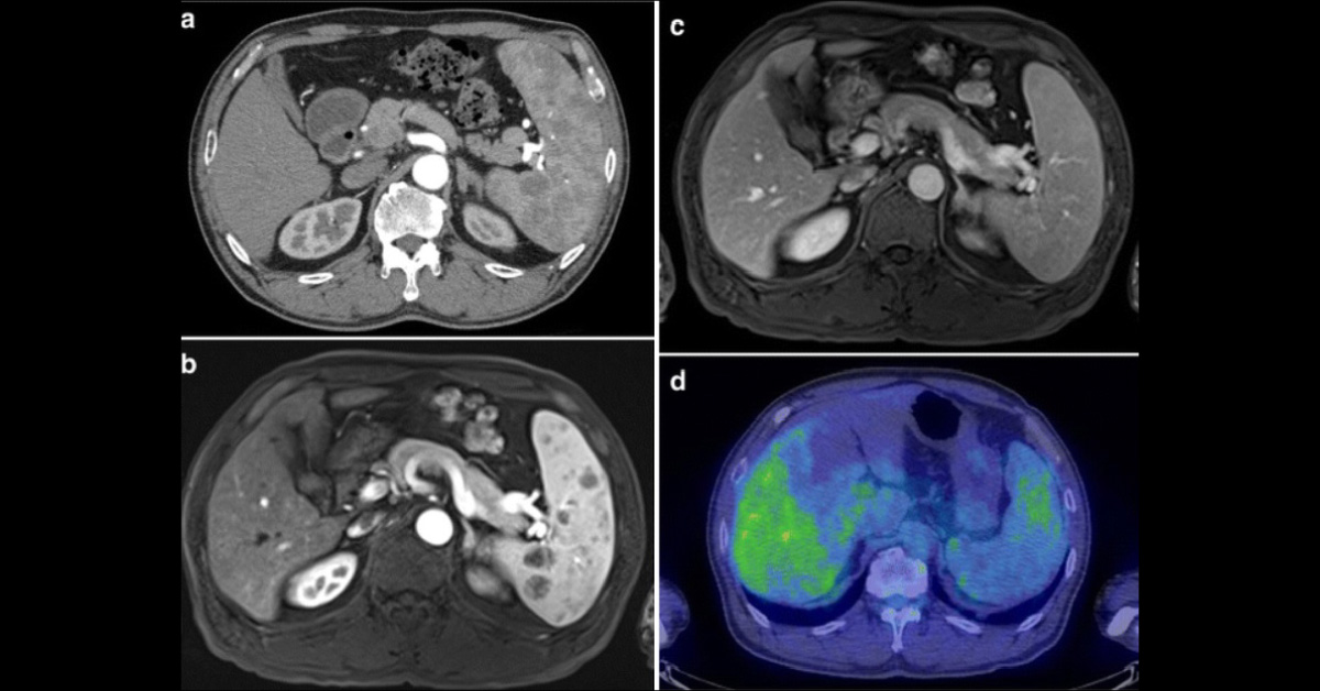
A 63-year-old man presented for a normal check-up but his ultra-sound revealed multiple splenic masses. He had a past history of hyperlipidemia. He had a BMI of 24.9 kg/m 2 . His triglyceride level was 963 mg/dL and total cholesterol 112 mg/dL. His LDL levels were 15 mg/dL, while HDL levels were 16 mg/dL. Plasma-soluble IL-2 receptor level was normal.
AUS demonstrated splenomegaly (spleen measurements 15 × 8 cm). Splenic echotexture was normal and multiple hyperechoic nodules were observed scattered throughout the spleen. Doppler imaging showed uneven blood flow in the lesion. Contrast-enhanced computed tomography (a), magnetic resonance imaging (b), dynamic MRI (c), positron emission tomography-CT assessment (d) has been provided. Preoperative images suggested extramedullary hematopoiesis, hamartoma or inflammatory pseudotumor. Differential diagnosis also considered metastatic splenic…



