
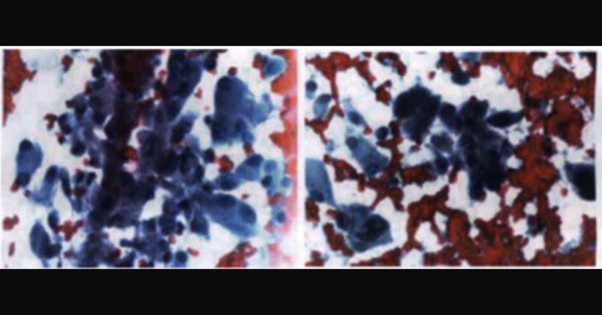
CLINICAL HISTORY A 70-year-old female was referred to a head and neck surgeon for evaluation of a left thyroid nodule. On ultrasound, the nodule measured 2.3 cm with calcifications. She subsequently underwent fine needle aspiration biopsy and specimens were sent for cytology, the result of which prompted molecular testing on the FNA specimen. CYTOLOGY (per outside report) The Papanicolaou stained direct smears were described as showing Hurthle cells characterized by uniform nuclei, prominent nucleoli, and abundant granular cytoplasm.
(See the Image) These were dispersed singly and in clusters in a hemorrhagic background. Binucleate forms were also seen. Molecular testing showed a RET p.C630R mutation (37% allele frequency) and high calcitonin gene expression What is your diagnosis? *This case is from Docplexus editorial team for educative purpose only Source: UPMC life-changing medicine…





