
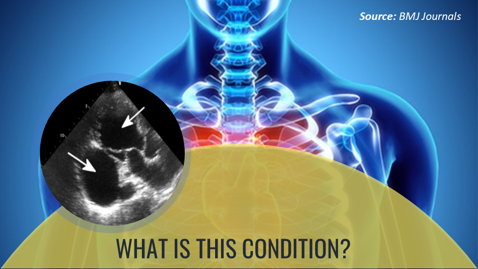
Case presentation A 69-year-old male reported to OPD after an episode of chest pain. Medical history No significant medical or trauma history Physical examination No pain at the time of OPD Hemodynamically stable On auscultation – Pan-systolic murmur was heard Electrocardiogram was normal Diagnostic tests Outpatient treadmill testing Transthoracic electrocardiography (Cover image) o Severely thin basal posterior wall of the left ventricle o Akinesis of the posterior wall o An outpouch adjacent to the posterior wall, measuring 4.5×7.5 cm 2 Color and pulse wave doppler test – Flow across the posterior wall (Figure 1) Figure 1.
Transthoracic ECG with color Doppler demonstrating flow across the posterior wall. (Source: BMJ Journals) Intraoperative findings o Three vessel coronary artery disease (CAD) o Severe stenosis of the mid-circumflex artery Based on the above…