
Abstract The aim of this study is to extract cortical and trabecular features of the mandible and to develop a novel combinational model of mandibular cortical thickness, trabecular bone area and age in order to predict low bone mineral density or osteoporosis from a dental panoramic radiograph. The study involved 64 south Indian women (age = 52.5 6 12.7 years) categorised into two groups (normal and low bone mineral density) based on total femur bone mineral density. The dental panoramic radiographs were obtained by a digital scanner, and measurement of total bone mineral density at the right femur was performed by a dual-energy X-ray http://pih.sagepub.com/content/early/2014/08/31/0954411914548700 absorptiometry scanner.
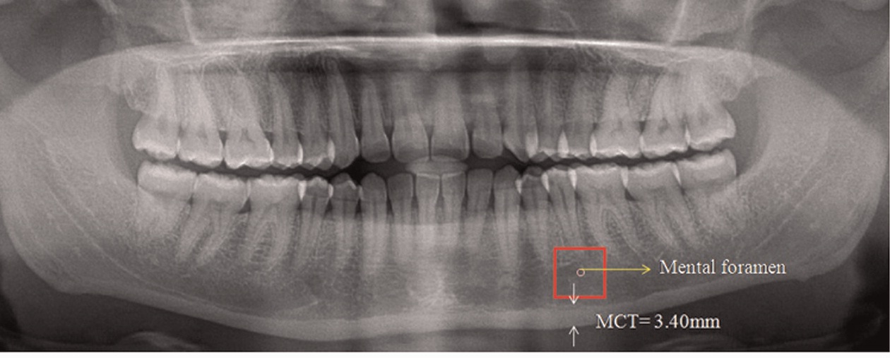
The mandibular cortical thickness and panoramic mandibular index were measured bilaterally, and the mean values were considered. The region of interest of 128 3 128…





