
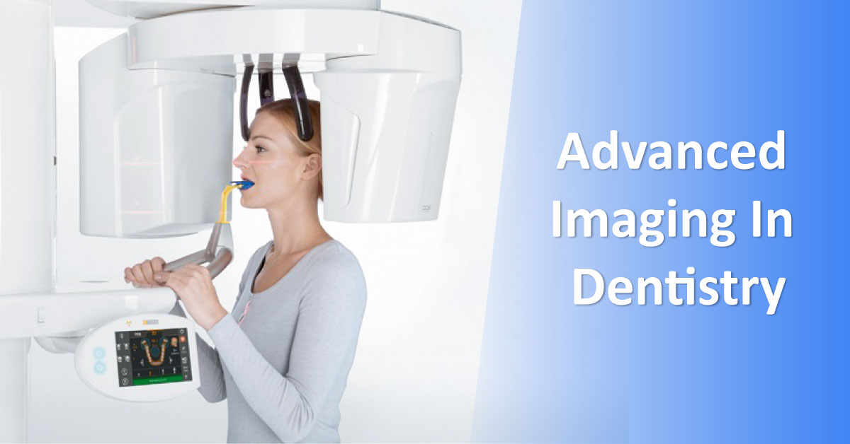
Radiographs are a valuable tool- in addition to clinical examination- for diagnosis of dental diseases. Although two dimensional (2-D) periapical and panoramic radiographs are routinely used in dental practice, there are certain limitations of these imaging modalities that can be overcome by 3-D imaging techniques such as cone beam computed tomography. Cone beam computed tomography, is based on a cone-shaped X-ray beam centered on a 2-D detector. It performs one rotation around the object and produces a series of 2-D images which are reconstructed in 3-D.
The major advantage of CBCT over CT is the considerably lower effective radiation dose to which patients are exposed. According to several reports, the radiation dose of one CBCT scan may be as little as 3-20% that of a conventional CT scan, depending on the equipment used and the area scanned. CBCT does not require an additional…





