
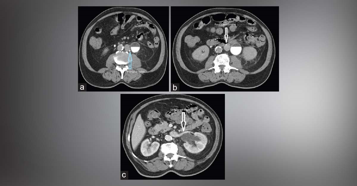
An 84-year-old male patient was presented to with dull, aching left flank pain of 6 months duration. He was moderately built, well-nourished and ambulant with no physical abnormalities. Ultrasonogram of the abdomen revealed left hydroureteronephrosis. X-ray of kidney, ureter, and bladder was non-contributory.
CT scan of the abdomen revealed transposed IVC to the left side and classic retrocaval ureter with proximal hydro-ureteronephrosis (images attached). All other viscera were orthotopically placed. Chest X-ray showed normally placed cardiac shadow. This case is form Docplexus editorial team for educative purpose only *Source: Cases in Urology Stay connected, the answer will be posted in 48 hrs!