
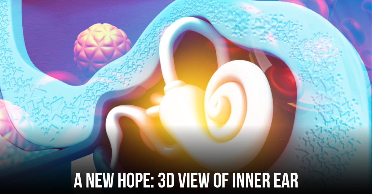
The inner part of the ear is protected by the temporal bone’s petrous side. The presence of this hard bone makes it difficult to study the anatomy of the inner ear. Recently, researchers from Uppsala University in Sweden have made it possible to depict inner ear details in a three-dimensional view. The study conducted by researchers from Uppsala University was published in Scientific Reports.
It highlighted the need for a better understanding of the micro-anatomy of the human auditory organ and has presented a three-dimensional (3D) reconstruction of the inner ear. The study also analyzed the human spiral ganglion (HSG) cell bodies blood supply using synchrotron radiation phase-contrast imaging (SR-PCI), combined with histological examination of the human cochlear sections. The synchrotron X-ray imaging is one of the eight systems in the world that use high energy accelerated particles…





