
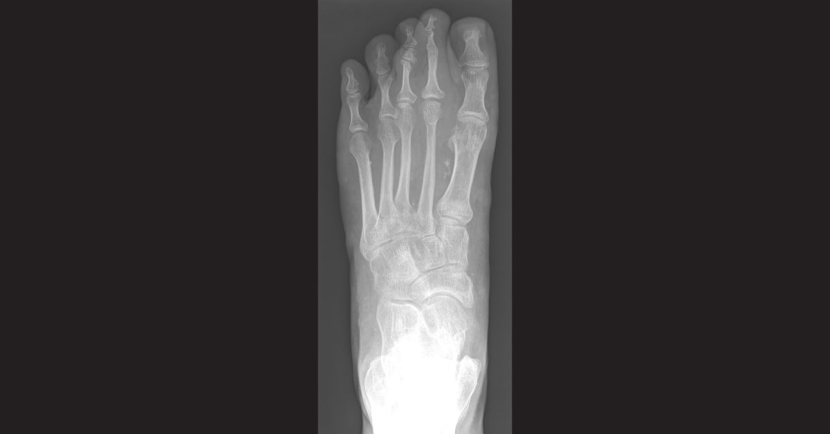
A 24-year-old male retired soldier presented with a 5-year history of a non-healing, painful left foot ulcer. He had a history of osteomyelitis requiring antibiotics, multiple debridements, and skin grafting. He underwent surgical intervention and endovascular treatment of an arteriovenous malformation (AVM) of the left lower extremity. Physical examination revealed a 6×2×0.2 cm ulcer on the dorsum of the left foot at the level of the second metatarsal head.
The dorsalis pedis and posterior tibial pulses of the left foot were palpable. Radiological findings Radiographs show clustered vascular calcifications between the first and second metatarsals, ankylosis of the second distal interphalangeal joint, and chronic bony remodeling along the diaphysis of the second middle phalanx, compatible with a slow-growing, benign adjacent soft tissue process (images attached). CT angiograms…





