
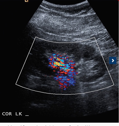
Case: A 57-year-old female presents with recurrent urinary tract infections and hematuria. She reports intermittent left flank and pelvic pain, which she has had for several months. Past medical history of hyperlipidemia and osteoarthritis. Past surgical history includes cystoscopy and hysterectomy. No family history of recurrent urinary tract infections or hematuria.
Physical exam of the patient was non-contributory, and the only lab value of significance was a serum creatinine 0.6 mg/dL. Imaging Findings: Ultrasound shows a left upper pole renal arteriovenous malformation, which measures approximately 2.6cm in diameter (Image 1). CT urogram shows a lobular mass within the left upper pole kidney, which was indeterminate but suspicious for vascular malformation (Image 2). Left renal arteriogram shows a large cirsoid type renal arteriovenous malformation. This occupies the majority of…