
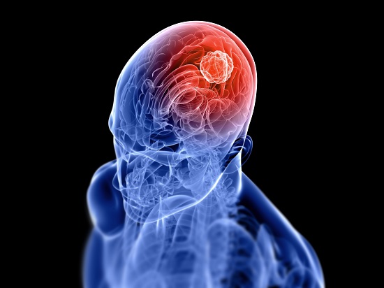
Till today, the only sources available to obtain data on the blood vessels in patients with tumors is either by taking a biopsy, a harmful procedure and often cannot be repeated, or PET scanning, which provided limited information and exposes patients to a dose of radiation. However, this information can be acquired in less two minutes in a single MR exam and can be safely repeated several times, through vessel architectural imaging (VAI). Traditional MRI, is a valuable technique to calibrate measurement of blood vessels for in vivo monitoring of hemodynamics status and vascular development, especially in the brain.
However, this technique as a limitation which is baffled by heterogeneous vessel architecture with stochastic perfusion. Which raised a question, how to identify a tumor response to anti-angiogenic therapy? Recently, a new imaging technique has made it possible to predict…





