
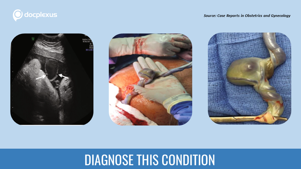
Case presentation At 30 weeks and 3 days gestation, a 38-year-old gravida 2, para 1001 woman was brought in from another hospital for managing reverse end-diastolic flow, as seen on umbilical artery Doppler assessment, along with elevated ductus venosus doppler. The fetal growth was restricted for the gestational age, less than the 3rd percentile. Medical history She had a cesarean delivery previously. The first-trimester screen had detected a high risk for trisomy 21 (1:80).
Type 2 diabetes mellitus Before the transfer to the present hospital, the patient had received two doses of betamethasone and a magnesium sulfate infusion. Doppler findings The previously identified findings of elevated ductus venosus with the reverse end-diastolic flow were confirmed. A 3-4 cm thin-walled, non-pulsatile lesion with a turbulent flow was detected in the free-floating umbilical cord. (Cover image)…





