
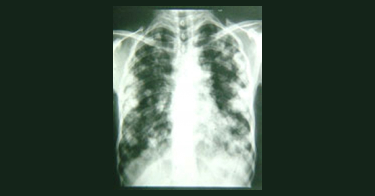
An- 8-year old boy was admitted for the second-time due to his progressive 10 day-long dyspnea. Two months back he was admitted to hospital hospital with clinical features of dyspnea, coughing and low-grade fever following a blunt chest trauma by a ball during his exercise. Two months back on his chest x-ray and computerized tomography scan, a large spherical lung cavity with a collapsed germinative layer was discovered in the right lung. According to the radiologic findings, operation was planned for the patient, presuming the diagnosis of a ruptured pulmonary hydatid cyst.
In the preoperative evaluation, the Casoni and indirect haemagglutination tests were negative and the liver and other abdominal organs were clear of infestation on ultrasonography. After two months, again the boy was suffering from dyspnea. What is the likely diagnosis based on X ray image attached? Source: Internet…





