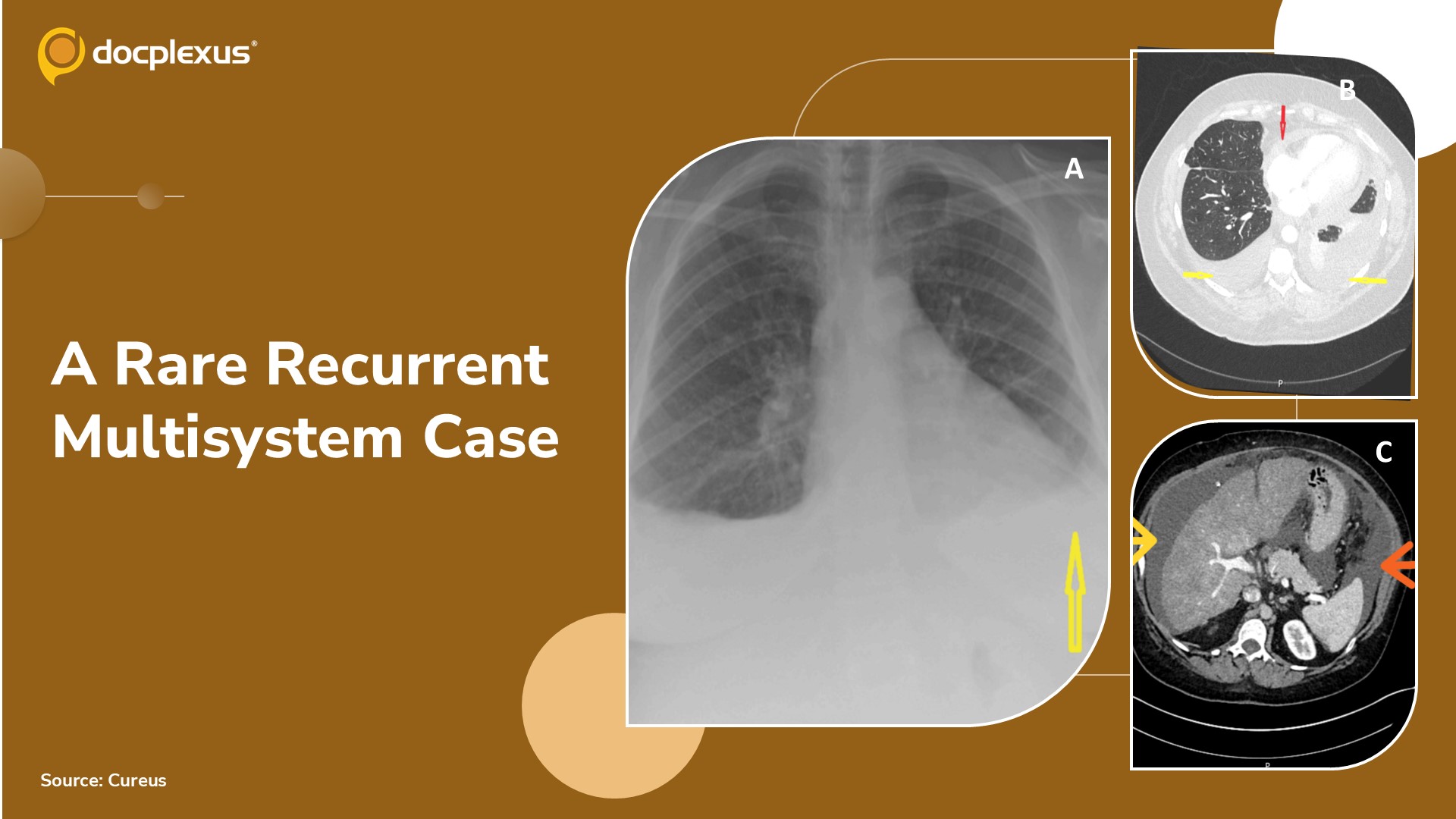
Case Presentation A woman in her 50s with no previously established diagnosis presented with recurrent symptoms requiring multiple hospital admissions. Her initial presentation was characterized by recurrent left-sided pleuritic chest pain accompanied by elevated inflammatory markers.
Based on these findings, she was initially treated empirically for suspected pneumonia. (Cover Image A) Early Investigations: CT pulmonary angiogram: No pulmonary embolism Bilateral pleural effusions Small pericardial effusion Managed as infection and discharged on antibiotics (Cover Image B) Subsequent Clinical Course Re-presented with: Progressive dyspnea on exertion Dry cough Bilateral pedal edema Abdominal distension (ascites) Generalized myalgia Required low-flow oxygen support Repeat Imaging Findings Persistent bilateral pleural effusions on chest X-ray CT thorax/abdomen/pelvis showed: Pleural…