
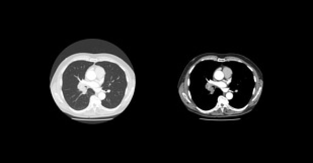
A 65-years-old male presented to the physician with shortness of breath and cough for a few weeks. Examinations: The CT scan are provided which shows the Axial lung window and the Axial C+ arterial phase. Further the patient was proceeded for bronchoscopy and biopsy. Microscopic Description: The sections show a proliferation of atypical lymphoid cells, forming abnormal follicular structures.
No diffuse areas were seen. Mixed centrocytes and centroblasts observed. What is the probable diagnosis? *This case is from Docplexus Editorial Team for educative purpose only Source: Radiopaedia Correct answer posted below ( Click Here )





