
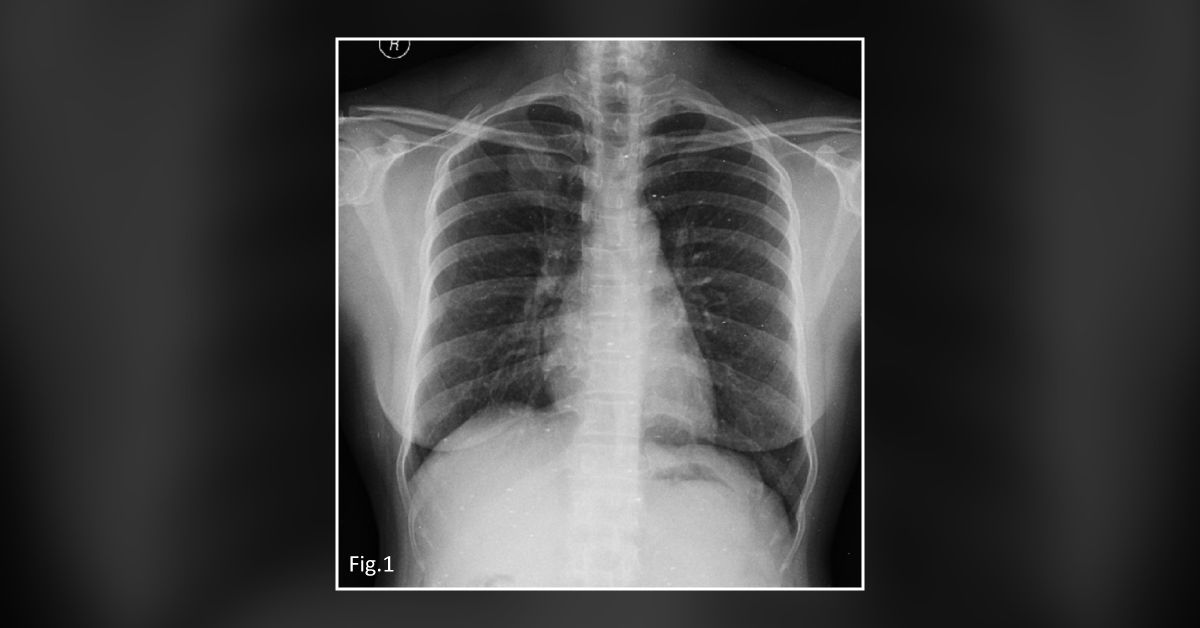
A 41-year-old female presented to a clinic with a 3-month history of cough. She did not report any history of hemoptysis or weight loss.
Clinical examination Normal physical examination Chest X-ray: A coin shadow in the right upper lobe ( Figure 1 ) CT scan: A homogenously enhancing spiculated soft tissue lesion in the apical segment of the right upper lobe (size 3.2 × 2.4 cm) Whole-body 18FDG positron emission tomography (PET)/CT: A high focal fluorodeoxyglucose-uptake by the lesion ( Figure 2 ) Fine-needle aspiration cytology: Inconclusive The patient underwent an upper lobectomy. The results are shown below: Gross examination: Partly circumscribed yellowish-white firm lesion, measuring 2.7 × 2.8 × 2.5 cm, with vague lobulation Section study: An ill-defined neoplasm with cellular as well as hypocellular areas Presence of spindle cells with elongated vesicular nuclei, small nucleoli…



