
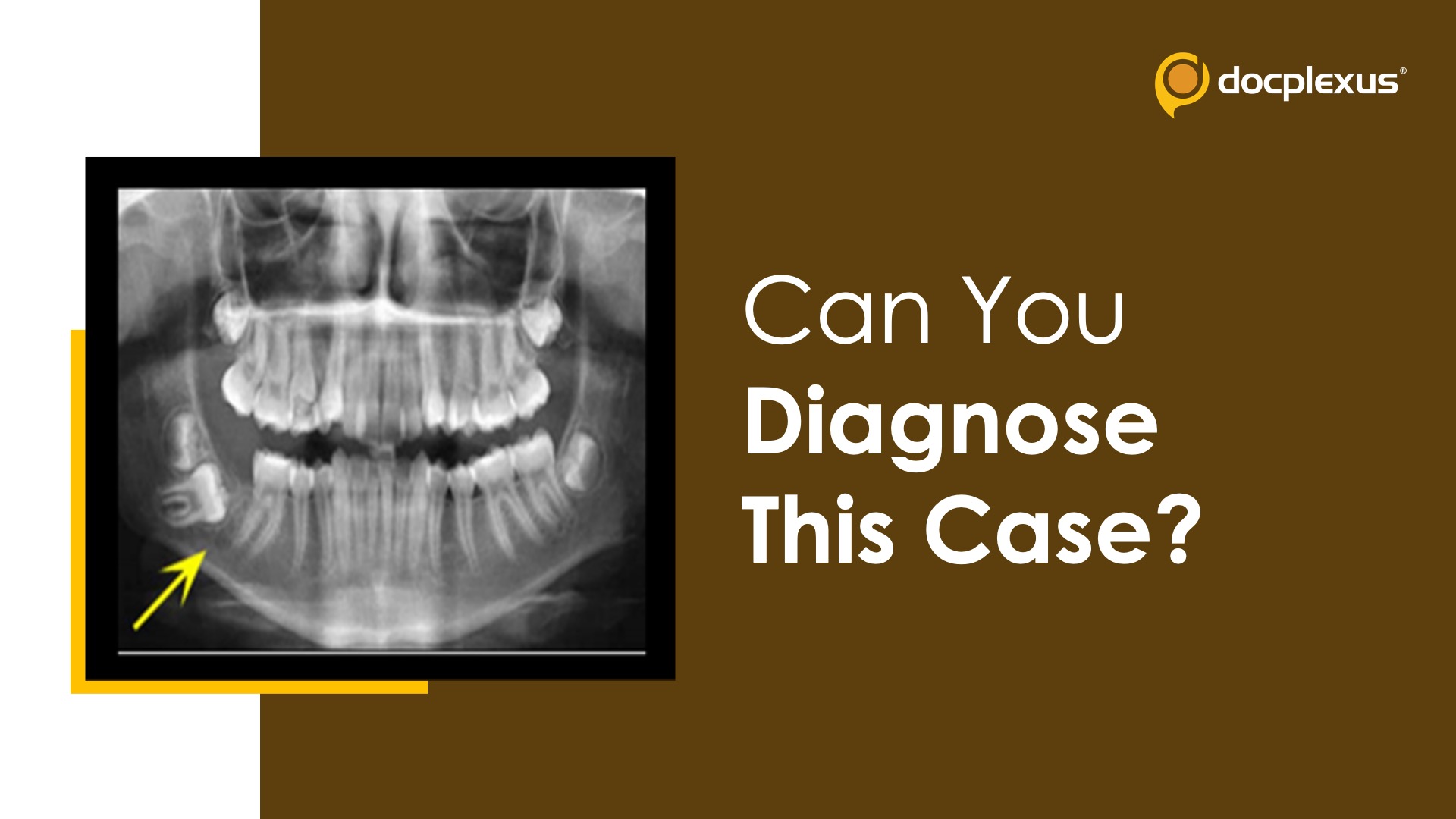
Case presentation: A 14-year-old female child with a tooth (31*) affected by a non-expansile radiolucent lesion was presented to the orthodontist. This lesion was an incidental finding. The patient was otherwise asymptomatic. Clinical history No allergies No significant medical or medication history Radiological investigations Panoramic radiograph: Unilocular radiolucency was found adjacent to the affected tooth. Cone-beam computer tomography (CT): A well-defined radiolucent area of 1.5cm was found, lingual and distal to the roots. Expansion and thinning of the lingual cortical plate were observed (Figure-1).
Figure 1 . Cone-beam CT. Source: Dental Care Histopathological findings (Figure-2) Several odontogenic nests in a myxomatous stroma The epithelium appears reminiscent of the developing enamel organ. Figure 2 . Biopsy findings. Source: Dental Care Source: Dental Care…




