
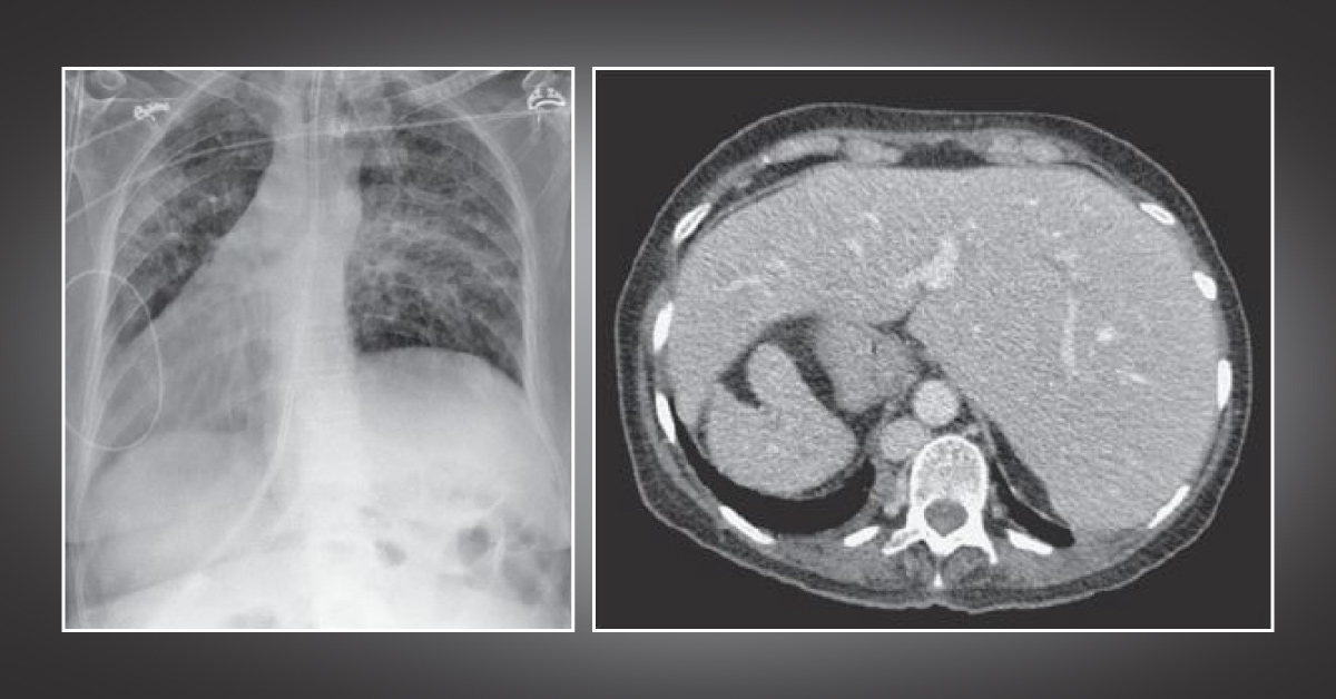
Case presentation A 41-year-old female was transferred from the general ward to the high-dependency unit because of deteriorating breathing. History Admitted with a short history of dyspnea and two days of coughing Demonstrated pyrexia and raised inflammation markers Treated with antibiotics and high-flow oxygen for presumed pneumonia, which continued to worsen No significant previous medical history Examination Tachypneic: 30/min Bronchial breathing in both mid-zones with inspiratory crackles in the same areas Air entry reduced bilaterally Heart sounds examination It cannot be appreciated in the normal left precordial position Apex beat is not palpable on the left Instead, the apex beat is palpated, and heart sounds are auscultated on the right of the precordium.
Palpation of the abdomen suggests a liver edge under the left costophrenic angle. The images of the chest radiograph and…





