
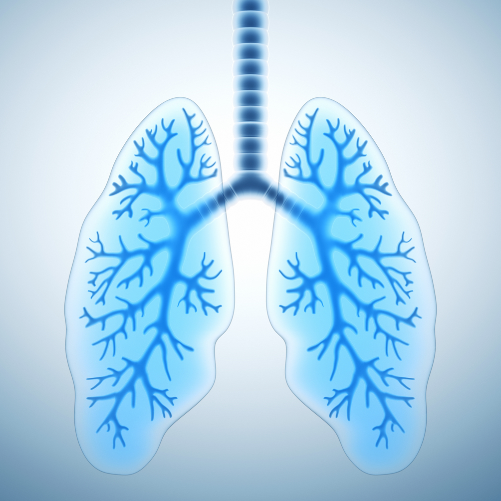
Recent procedures like computed tomography and improved three-dimensional (3-D) post-processing techniques have revolutionized the capability of visualizing airway pathology, offering physicians an advanced view of pathology and allowing for appropriate management planning. This article describes the imaging modalities used for trachea and main bronchi imaging. imaging of the respiratory tree or trachea-bronchial tree has enhanced due to the advancements of computed tomography (CT)) that permits volumetric isotropic voxel imaging.
It's associated post-processing software allows for advanced three-dimensional (3-D) visualization. Thus, airway imaging has become a tool that physicians can use to help plan interventional procedures, like bronchoscopy, stent placement, surgery and subsequent follow-up. Imaging modalities MDCT, including thin-slice volumetric CT, paired inspiratory and…





