
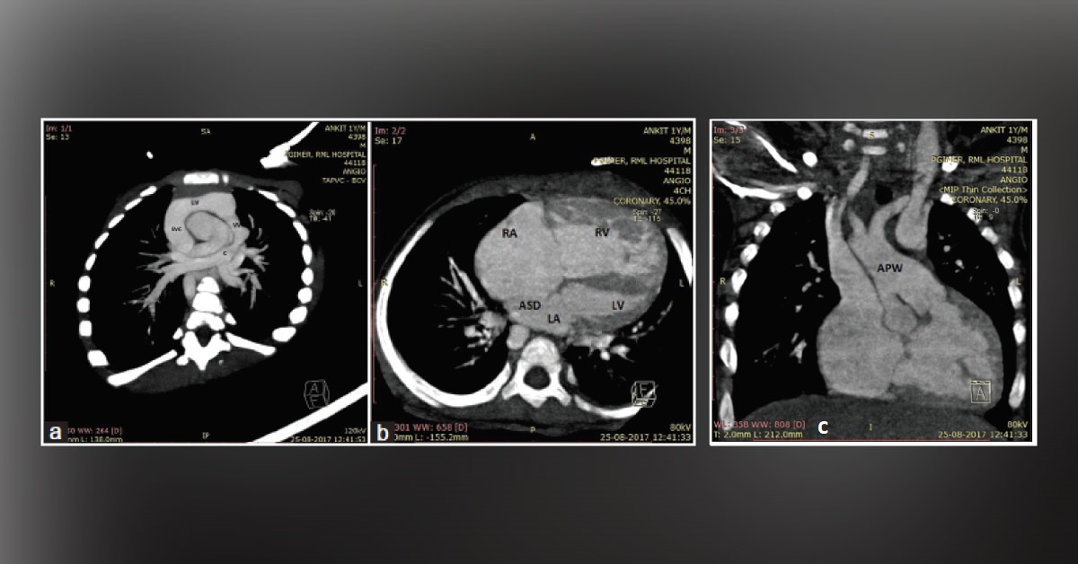
An 8‑month‑old male child was admitted with a history of frequent cough and respiratory distress from three months of his age. He was referred following a detection of a murmur by his pediatrician. At admission, he was in congestive cardiac failure. A Grade 3/6 short systolic murmur along the parasternal area with loud and palpable second heart sound was remarkable. Resting saturation was 85%.
Chest roentgenogram showed cardiomegaly and pulmonary plethora. Echocardiography showed enlarged right atrium, right ventricle (RV), pulmonary artery, supracardiac type TAPVC (three pulmonary veins were delineable) with an ostium secundum atrial septal defect (ASD) with a right to left shunt, and severe pulmonary hypertension. He was treated with oxygen and decongestive measures. Cardiac computed tomography (CT) was done to delineate the anatomy of pulmonary veins. It confirmed the presence of…





