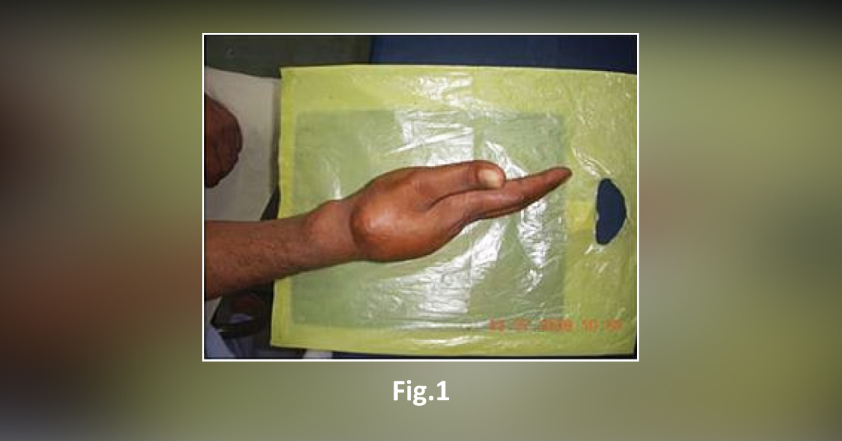
A 48-year-old man presented to an orthopedic outpatient department with a complaint of a painless mass in the right hand. The patient reported that the painless mass had been slowly increasing in size over the past several months. Physical examination A well-defined firm, nontender, nodular mass measuring 10 X 7 cm [Fig. 1] . Radiological finding X-ray of the right hand showed radio-opaque tumor-like mass with granular fluffy pattern obscuring the underlying bony details [Fig. 2] .
Cytopathological results Fine-needle aspiration cytology (FNAC) was performed using 23 G needle attached to a 10 ml syringe. FNA yielded thick, white, chalky, gritty material. Smears were made from the aspirate and stained with Giemsa. Light microscopy of air-dried, Giemsa-stained smears showed abundant calcified areas, histiocytes and rhomboid-shaped crystals [Fig. 3]. Histopathological analysis Biopsy…