
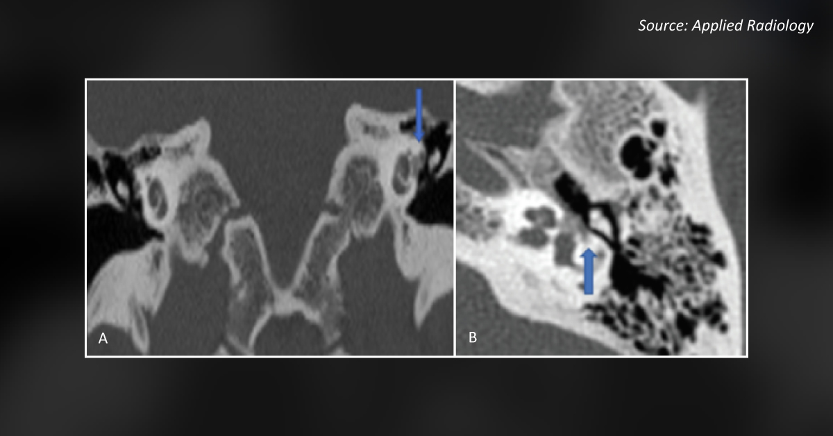
Case: A 57-year-old woman presented to the clinic with complaints of left-sided hearing loss, intermittent otalgia, and superimposed subjective non-pulsatile tinnitus. She had a history of progressive hearing loss which had worsened over the last year. An audiological examination was remarkable for mixed conductive and sensorineural hearing loss of the left ear. A CT scan of the temporal bone demonstrated resorptive changes along the margins of the oval window (Image A) and left cochlear promontory (Image B) with left capsular sparing on the right. Question: Based on the HRCT findings, can you diagnose the condition of this patient? A.
Meniere’s Disease B. Acoustic neuroma C. Fenestral otosclerosis D. Presbycusis Stay tuned for the right answer! *This image challenge is exclusively from the Docplexus Editorial team for an educative purpose only! Source: Applied Radiology