
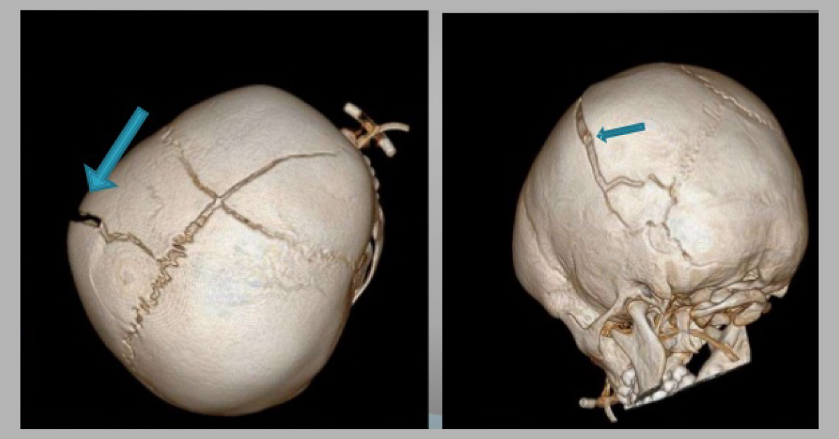
Plain film radiography remains the most cost-effective method for evaluating skull fractures and can easily differentiate major sutures and common vascular grooves from fractures. However, in children, this can be complicated due to the presence of numerous synchondroses and unusual accessory sutures. The current article outlines the radiological differentiations that can aid in the identification of skull fracture from accessory sutures in pediatric patients.
Challenges with plain film radiography Plain film evaluation is especially challenging not only because of various artifacts that can degrade the study but also the inability to visualize intracranial processes, such as contusions and hemorrhage, that can substantiate a calvarial finding. Minimal soft tissue swelling can be difficult to see even with oblique views. Superimposition of normal suture lines like the metopic suture can…





