
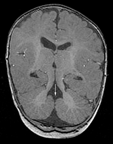
A 3-month-old male infant was initially diagnosed with a Dandy-Walker malformation based on head CT findings. He was born by C-section 3 weeks prematurely due to rupture of membranes and was in the neonatal intensive care unit for several weeks after birth due to poor feeding and episodes of tachypnea. He was brought to the emergency room for evaluation of increasing lethargy and increased head size. The clinical examination revealed nystagmus with abnormal eye movements, small optic discs, frontal bossing, upturned nasal tip, recurrent episodes of hyperpnoea and developmental delay.
Because his clinical examination could not be solely attributed to a Dandy-Walker malformation, an MRI evaluation was performed (images attached). Imaging findings The MRI revealed an abnormal appearance of the posterior fossa. A severe hypogenesis of the cerebellar vermis, enlarged, horizontal superior…





