
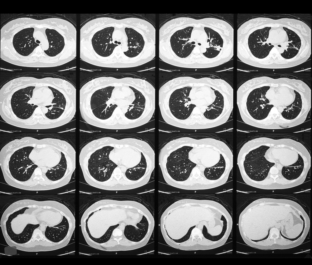
High-Resolution Computed Tomography (HRCT) is a widely used technique to image various lung pathology. HRCT uses a narrow beam collimation to take thin slice images of the lung parenchyma. This protocol produces extremely high definition images of lung alveoli, airways, interstitium, and pulmonary vasculature. Expiration images may assist in identifying air-trapping in patients with lung disease. The initial imaging tool for the lung parenchyma is chest radiograph.
It is unexcelled in the information it yields in relation to its cost, a dose of radiation, availability, and performance. Due to some limitations of chest radiographs in several studies, chest radiographs are mostly not preferred. For these reasons, high-resolution computed tomography (HRCT, also called thin-section CT scanning), is frequently used to help clarify specific problems. HRCT High-resolution computed tomography…





