
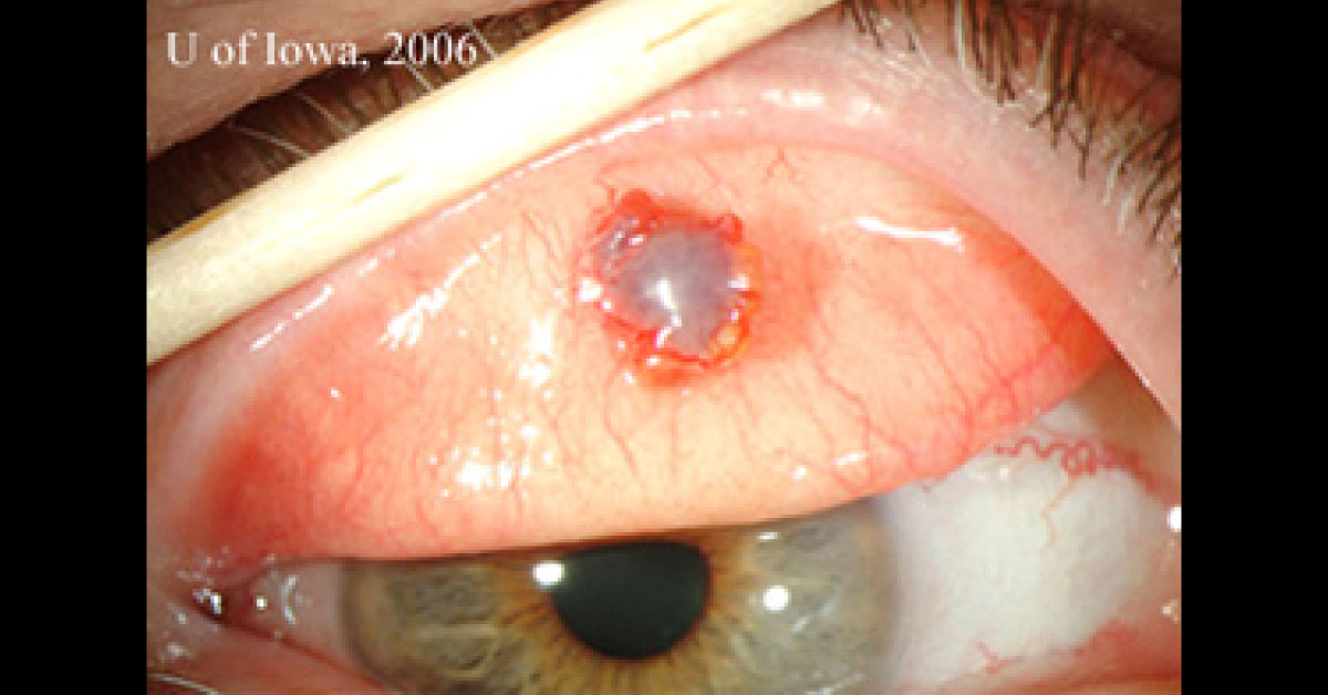
Patient History: The patient is a 67-year-old male presents with a pigmented conjunctival lesion in his right upper eyelid. He initially noticed it 1 year ago but was asymptomatic until 6 months ago when he started experiencing decreased visual acuity and a foreign body sensation in that eye. He initially thought that his symptoms were due to dry eye.
Physical Examination: Eversion of the right upper eyelid reveals Erythematous, elevated lesion within the tarsus of the right eyelid and protruding through the upper palpebral conjunctiva of the right eyelid (image attached). H&E stained section of the first eyelid wedge specimen reveals atypical dark, pleomorphic sebaceous cells (black arrows) which are in marked contrast to the normal foamy sebaceous glands (white arrows) seen in the upper field of the photomicrograph (image attached). What is a possible medical diagnosis in this case?…