
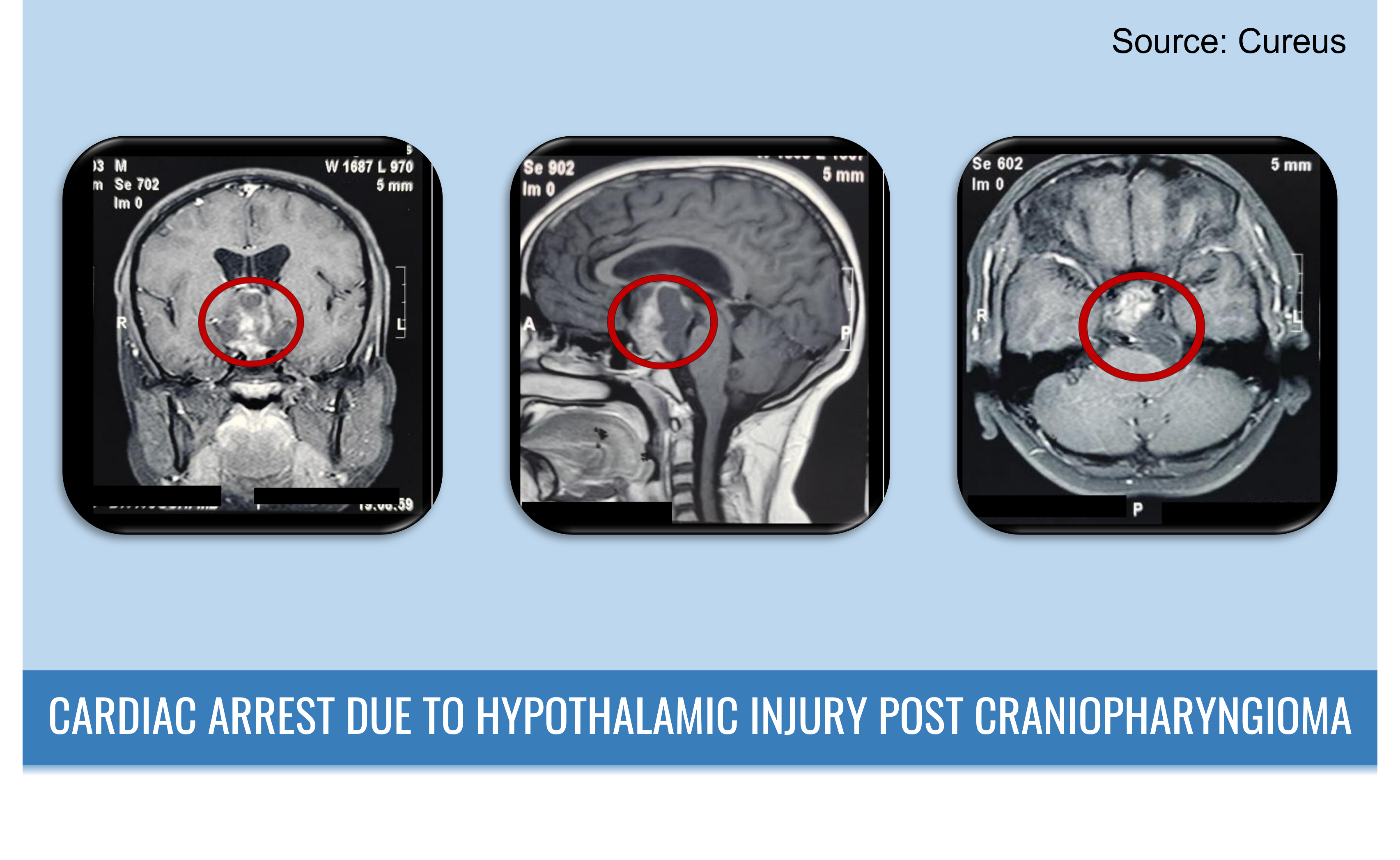
Case presentation A 13-year-old male patient was hospitalized with complaints of recurrent seizures and an eight-month history of headaches. Clinical examination Neurological examination was unremarkable and showed no motor or sensory deficits. Bilateral papilledema was observed. Radiographic examination A multicompartmental tumor was noted in the MRI, extending from the suprasellar cistern to the right subtemporal area, infiltrating the prepontine cistern, and weakly adhering to the basilar artery.
Figure 1. Preoperative MRI scan. (Red circle marks the location of the lesion on the MRI) Surgical management A left frontotemporal craniotomy, also known as pterional craniotomy, was performed under the influence of antibiotics, anti-epileptics, and mannitol. The arachnoid matter was opened with the help of a transsylvian approach, and the right internal carotid artery and optic nerve…

