
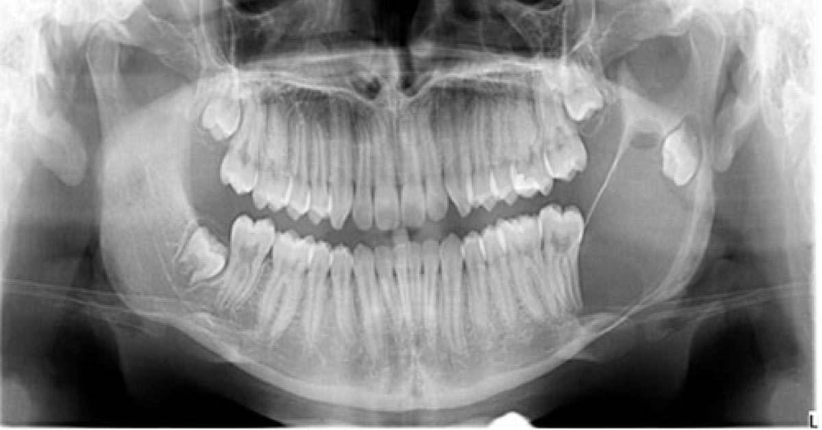
During a routine check-up of a 16-year old boy, his dentist noted a left mandibular swelling. A panoramic radiograph revealed a very large and well demarcated unilocular radiolucency associated with the crown impacted tooth. Bony perforations were noted in the ramus area. The expansion was intra-oral, mild, and more buccal. Histology revealed a cystic structure lined by keratinized epithelium and supported by a cellular connective tissue wall.
The connective tissues were is cellular and infiltrated by lymphocytes and plasma cells. Identify this condition in this young boy. *This case is from Docplexus editorial team for educative purpose only. Source: NEJM Stay connected, the answer will be posted within next 48h!