
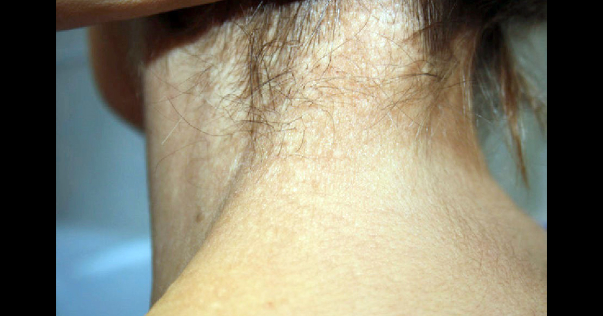
Medical History : A 65-year-old woman with a 13-year-history of primary biliary cirrhosis, systemic hypertension, dyslipidemia, and osteoporosis came to our clinic for evaluation of lesions that had developed 2 years earlier. The lesions had gradually appeared on the back of the neck and were asymptomatic. Physical Examination: E xamination revealed multiple whitish papules measuring 3 mm in diameter and with a rubbery consistency; the follicular openings were spared and showed no mucinous material when pressed. The papules appeared in clusters on the back of the neck (image attached), with occasional papules in the right axilla.
Additional Tests: Dermoscopy revealed well-delimited, homogenous, whitish papules with some dotted vessels at the periphery. A 4-mm punch biopsy taken from a lesion on the neck showed slightly thickened collagen fibers in the mid dermis (image attached). No…





