
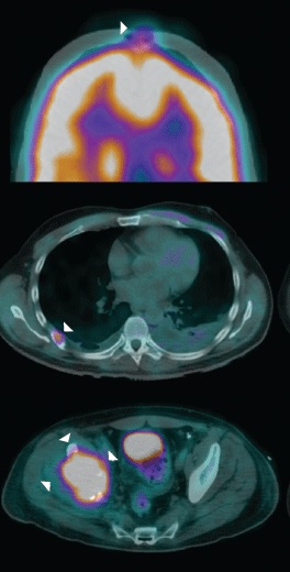
A 42-year-old man presented with increasing right hip pain that limited his ability to walk. Imaging Studies Magnetic resonance imaging (MRI) revealed a right lytic acetabular lesion. Further work-up included a computed tomography (CT) scan, which revealed an 8-cm left kidney tumor, and a positron emission tomography (PET)/CT scan (Image 1), which showed osseous metastases involving the right acetabulum, the posterior end of the sixth right rib, and the cranium (frontal bone). Surgical management and Pathology The patient underwent a left open radical nephrectomy.
The pathology evaluation revealed an 8-cm papillary type 2 renal cell carcinoma, with scant areas of clear-cell renal cell carcinoma, and negative margins (Image 2). He made a good recovery from surgery and completed palliative stereotactic body radiation therapy to the right acetabular lesion. What is the staging of his…





