
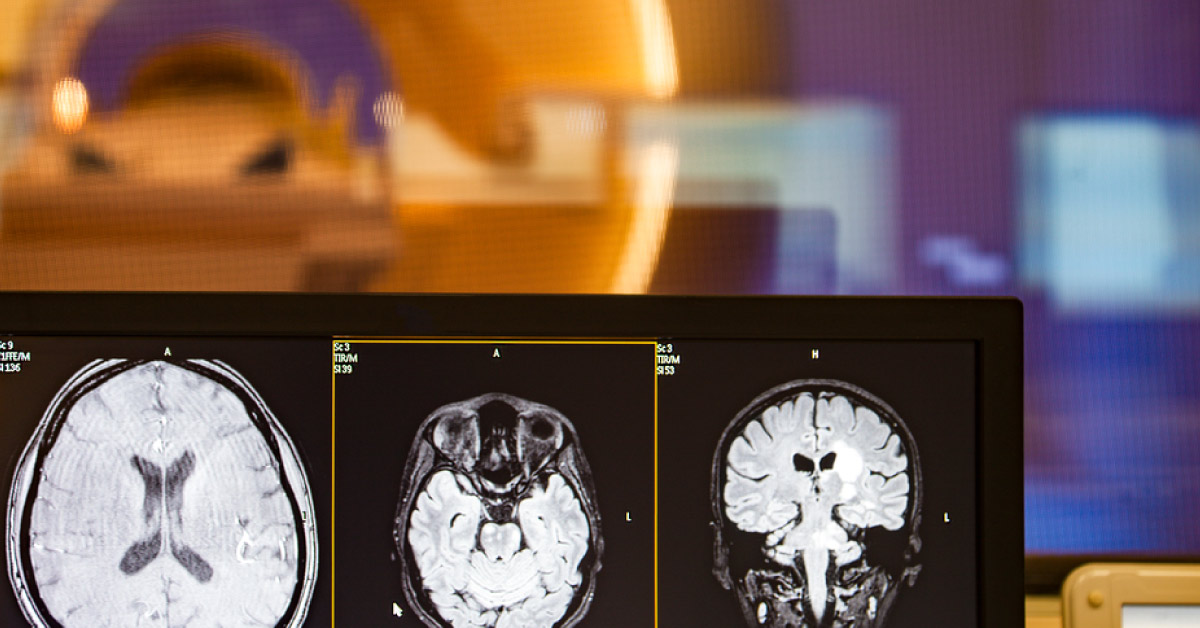
The use of natural, unlabeled D-glucose as a biodegradable contrast agent to image glucose uptake and, potentially, glucose metabolism in vivo has been demonstrated in animal tumor models. The current article cites the study that performed T1r-weighted DGE MR imaging at 7.0 T in healthy volunteers and patients with newly diagnosed, untreated glioblastoma to evaluate the potential to detect intracerebral regions of increased glucose concentration during intravenous glucose injection. The concept used : In magnetic resonance imaging (MRI), contrast agents are used to enhance the imaging of tissue structures.
While they enhance signals in blood vessels and in spaces between cells, they do not reach the interior of the cell. By contrast, glucose is taken up and then broken down in the body cells – Warburg’s effect. Tumor cells are particularly hungry for glucose in order to feed their high…





