
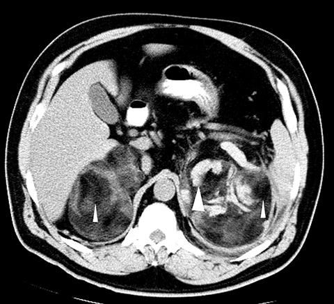
A 50-year-old male presented with two days of left flank pain associated with nausea and vomiting. Physical exam revealed left flank tenderness; however, serum chemistries and CBC were within normal limits. Abdominal CT showed bilateral adrenal masses, left larger than right. Surgical resection of the lesions yielded well-circumscribed specimens with smooth capsules and a heterogenous internal composition consisting of adipose tissue and blood products.
The patient experienced an uneventful recovery. Imaging findings Computed tomography was done (Images attached) What diagnosis can be inferred from the radiological findings for this patient? *This case is from the Docplexus Editorial Team and is meant for educative purpose only.