
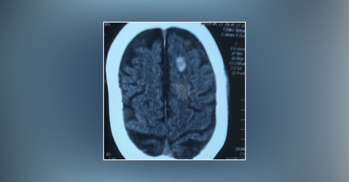
Recently a man of 80 years had some form of unconsciousness and then recovered. CT scan taken in a medical college hospital where he was admitted showed a possible intracerebral haemorrhage in the left frontal region and is very small, as per the Imaging Specialist. He was treated accordingly but the patient was alright with no other neurological symptoms and signs. He was advised to have an MRI scan and was taken. The MRI scan reported as Subacute infarct by another Imaging specialist.
The patient was admitted in the ward of another government hospital and on seeing both which showed the same finding namely opaque elliptical lesion in both images. As per the literature when the lesion is opaque in CT Brain it is intracerebral haemorrhage if the vascular lesion is thought of. In the same way, in MRI it is infarct. But one will have to see the lesion fully namely its border, shape and…





