
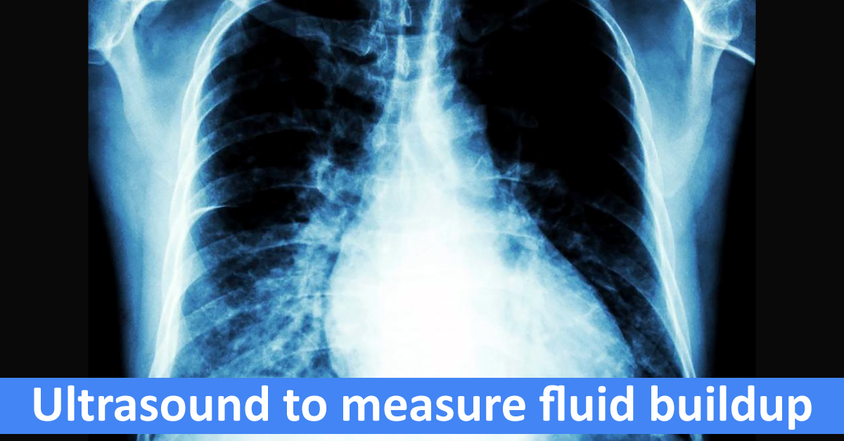
A team of researchers have found a noninvasive way to use ultrasound to monitor fluid levels in the lung, offering to track progress in treating pulmonary edema – fluid in the lungs – which often occurs in patients with congestive heart failure. The approach, holds promise for diagnosing scarring, or fibrosis, in the lung. Challenges and shortcomings of the conventional method Conventional lung imaging is generally done using chest radiography (CXR) or thoracic computed tomography. Both these imaging modalities have limitations, which places constraints on their applicability.
CXR is constrained by limited diagnostic performance, portability of bedside radiography and X-ray exposure issues. Because of the moving thorax, the spatial resolution decreases and leads to poor quality X-ray films with low sensitivity. X-ray beam origination is not tangential to the diaphragmatic cupola,…





