
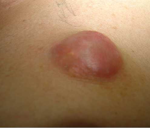
Case: A 56-year-old woman presented to the breast imaging centre with a several-month history of an enlarging, superficial, firm and non-tender palpable lump in the left breast at 12:00 (Image 1). The patient was post-menopausal, had no personal history of any type of cancer, and had no family history of breast cancer. Diagnostic mammography and breast ultrasound were performed for further evaluation of the palpable lump.
Imaging Findings: Mammography in the craniocaudad and mediolateral oblique planes demonstrated a well-circumscribed, equal-density oval mass measuring approximately 2.5 cm at 12:00 in the left breast at the site of the palpable lump indicated by a metallic BB marker (Image 2). Ultrasound revealed a circumscribed heterogeneous dermal mass with posterior acoustic shadowing and marked peripheral color flow measuring 2.6 cm. There was an abrupt interface with the normal…





