
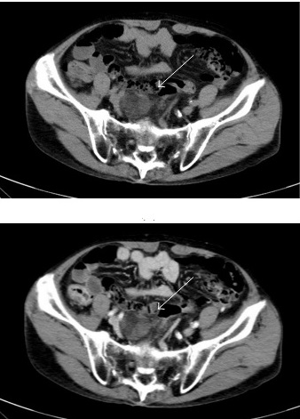
A 71-year-old male underwent abdominal computed tomography (CT) as part of the first follow-up of an intraductal papillary mucinous neoplasm of the pancreas. The patient had no remarkable symptoms. His medical history included alcoholic liver disease.
On physical examination, his abdomen was soft, and no palpable masses were detected.There were no significant signs of sacral root involvement, such as defecation or urination disorders, and there were no abnormalities in the patient’s laboratory data. Contrast-enhanced CT was performed, which revealed a heterogenous mass in the presacral space (diameter: 43 mm) that contained low-density areas suggestive of fatty components and some slightly enhanced areas suggestive of soft tissue islands inside the mass (image attached). Subsequent pelvic magnetic resonance imaging (MRI) showed a 43 mm mass in the anterior aspect of the sacrum (image…





