
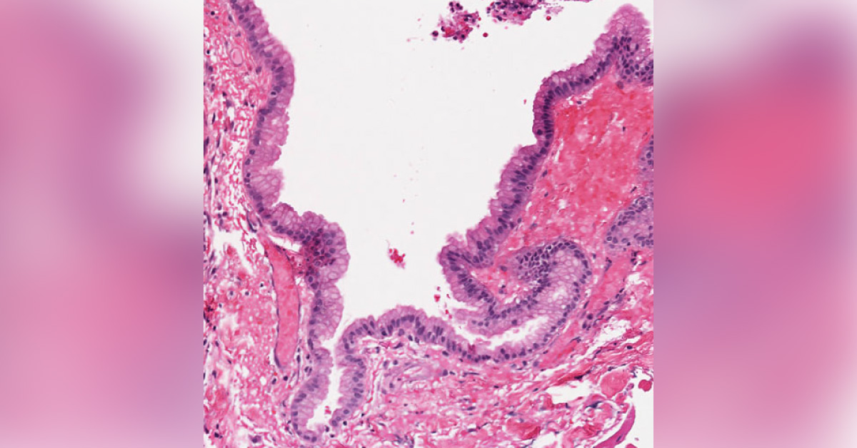
A preadolescent female is presented to the physician with nausea, vomiting, blurred vision, diplopia, gait instability and ataxia. An MRI showed a 22 x 21 x 78 mm enhancing tumor with a necrotic center located at the cranio-cervical junction expanding the medulla oblongata and the proximal aspect of the spinal cord to the level of T3. Additionally, there was a non-enhancing exophytic component of the tumor abutting the left pre-medullary cistern. The radiologic impression was an infiltrative, likely astrocytic, cranio-cervical junction tumor with an exophytic component.
Intraoperative Findings: The smear preparation showed proteinaceous debris and neutrophils which was interpreted as necrotic tissue. Microscopic Findings: Permanent sections showed membranous fibrovascular tissue lined by columnar to pseudostratified and partially ciliated epithelium. Proteinaceous mucinous material was…