
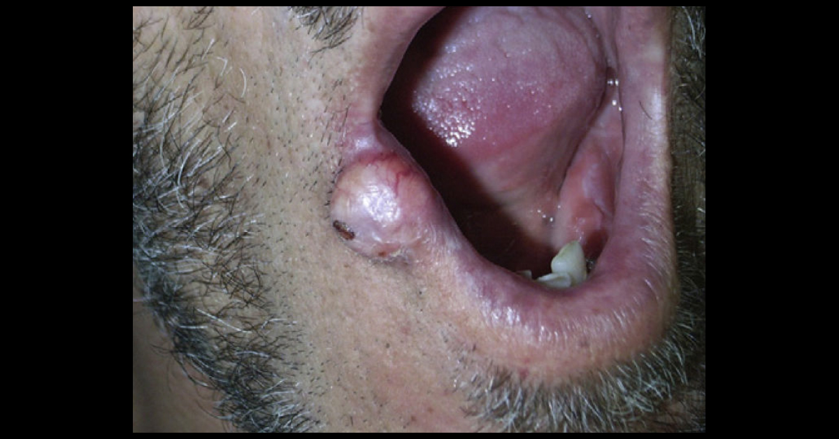
A 52-year old patient with no relevant past medical history presented with an asymptomatic, slow-growing, homogenous nodular lesion on the lower lip that had appeared 10 years earlier. The patient was unable to relate the onset of the lesion to any event and had received no previous treatment. Physical Examination: The patient had a well-defined pinkish-red nodule with superficial telangiectasia. The lesion infiltrated the full thickness of the lip, was painless on palpation and measured 1.5 cm in diameter (image attached).
Histopathology: Hematoxylin-eosin staining revealed the presence of lymphoid cell proliferation, consisting mainly of plasma cells in different stages of maturation, with the involvement of striated muscle tissue (image attached). Immunohistochemistry revealed κ light chain restriction but was negative for λ light chain and for CD20 (Image attached). What is the…





