
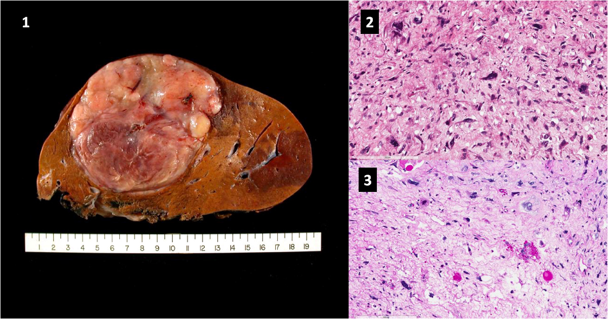
A 27-year-old female presented to the emergency room with intractable abdominal pain she had already experienced for several days. CT scan revealed a 14cm liver lesion in the right lobe (Image 1). The lesion appeared fluid-filled and had characteristics suggestive of a hemangioma. At the time of resection, intraoperative ultrasound suggested that lesion was solid. A right trisegmentectomy was performed. Pathologic Findings The tan-white mass had a gelatinous cut surface.
This lesion had a tendency to bulge from the cut surface and contained focal areas of central haemorrhage. The mass was lobulated at its periphery, with some lobules of lesion extending into the surrounding liver parenchyma. No other discrete lesions were identified. Histology The tumour was composed of haphazard arrangements to sheets of spindle, oval and stellate cells embedded in a myxoid stroma. In some areas,…





