
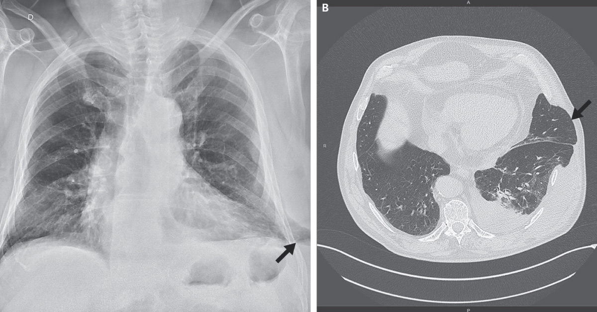
A 67-year-old man admitted to ED with a 10-day history of the painful left side of the chest that had begun after an episode of severe coughing. The pain was persistent without dyspnea that did not recede with analgesics. He also showed swelling on the lower life side of the chest. The patient had a history of smoking, hypertension, and chronic obstruction pulmonary disease (COPD) that requires continuous positive airway pressure. He denied any chest trauma or surgery involving chest.
Examination Physical examination revealed thoracic asymmetry, with a soft, reducible mass at the swelling that was painful on palpation. The mass increased in size on inspiration and decreases with expiration. Radiograph (image A) and CT (image B) were acquired. Spot the condition from the attached radiograph and CT as indicated by the arrow in the images. *This case is from Docplexus editorial team for…





