
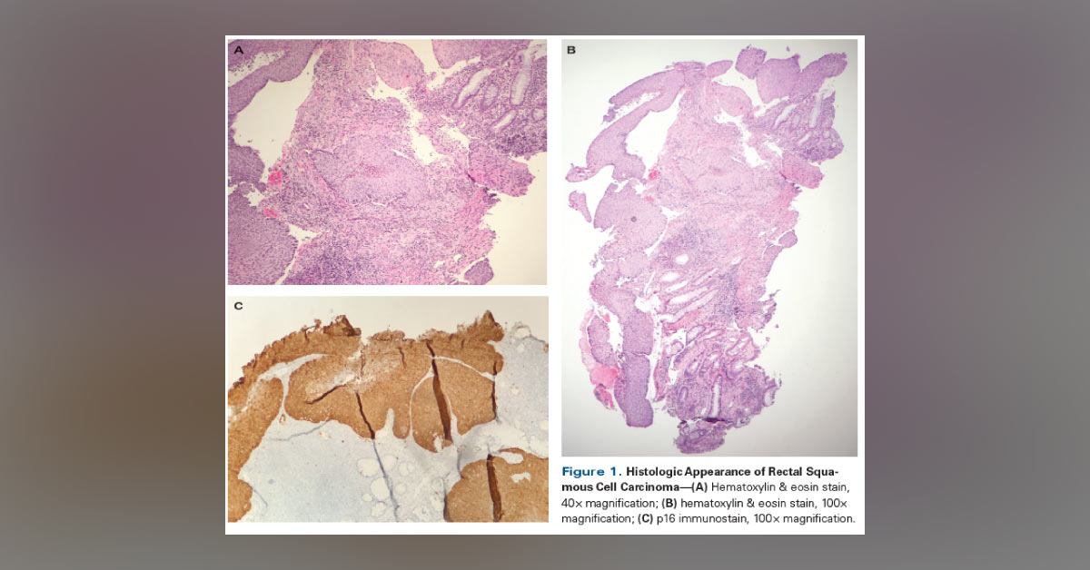
A 56-year-old woman presented to the clinic with a history of recurrent rectal blood with defecation for three months. At her initial visit, anoscopy and a pelvic examination for high-risk human papillomavirus (HPV) were performed, which showed negative results. A colonoscopy was also recommended, which revealed an abnormal area with a 3 × 4–cm mass in the rectum which was consistent with invasive squamous cell carcinoma (Figures 1A and 1B). Immunohistochemistry staining of the biopsy specimen was found positive (Figure 1C).
A total body CT scan (Figure 2A) and positron emission tomography (PET)/CT scan (Figure 2B) established the presence of a rectal mass. Any evidence of nodal or distant metastasis was absent. *This image-based case is from Docplexus Editorial Team for educative purposes only. Source: Cancernetwork- Home of the journal Oncology Answers are posted in the comment…





