
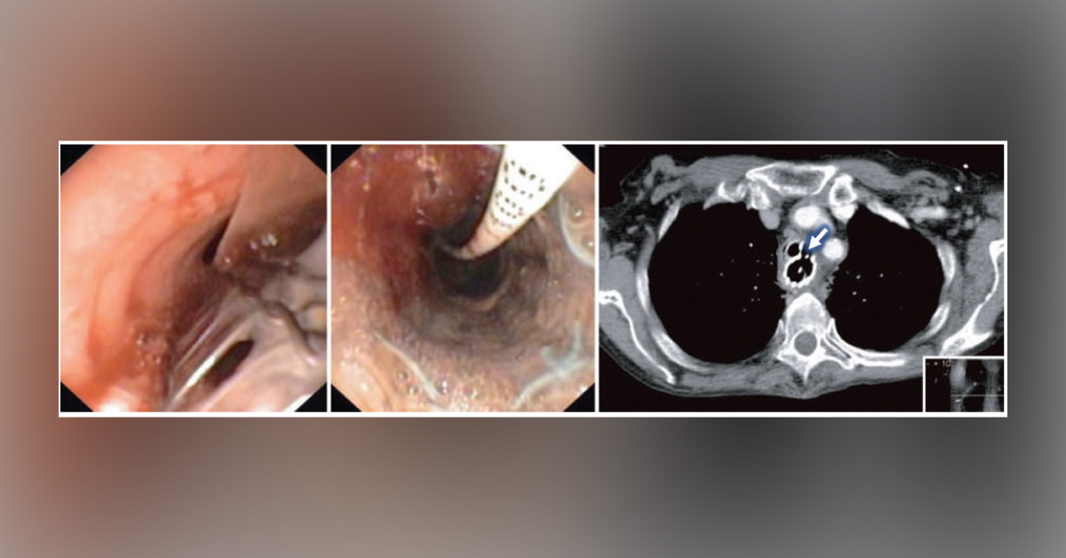
A 63-year-old lady was brought to the clinic with a complaint of dysphagia. The doctor went through her reports and came to know that the dysphagia might be a result of proximal esophageal stenosis, 27 years after she went through radiation therapy for Hodgkin's disease. The patient had experienced several other complications because of this treatment, including myopathy, cardiomyopathy, and hypothyroidism. Medical investigation Tracheobronchoscopy It revealed a long [6 cm] semi-circular defect in the trachea, beginning 2 cm carnia from the cranial.
The doctor also noticed the esophageal stent material bulging into the tracheal lumen. Esophagoscopy An esophagoscopy image revealed the lumen of the esophageal stent along with a feeding tube in situ . CT scan An axial image of the CT scan revealed a ventilation tube in the trachea and a stent in the esophagus, with complete…





