
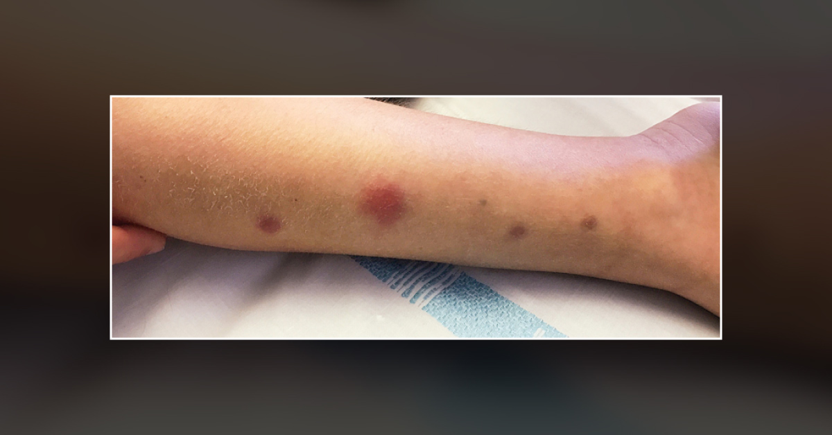
An 8-year-old boy presented to a dermatologist with subcutaneous nodules on the legs (as seen in the cover image) and one-week history of fever. The boy informed that the fever was persistent despite treatment with broad-spectrum intravenous antibiotics and antifungals. Additionally, he had been undergoing treatment for relapsing B-cell leukemia.
A physical examination revealed the presence of nodules measuring 5 to 18 mm in diameter on the chest, back, arms and legs. His laboratory workup was as follows: Hemoglobin – 7.5 g/dL White blood cell count – 200 per cubic meter Platelet count – 12,000 per cubic meter Following the laboratory examination, a biopsy of the nodules was performed and a microscopic examination was conducted using lactophenol cotton blue, which showed septate hyphae and spores. Source : The New England Journal of Medicine ##Disclaimer## The content provided on…


