
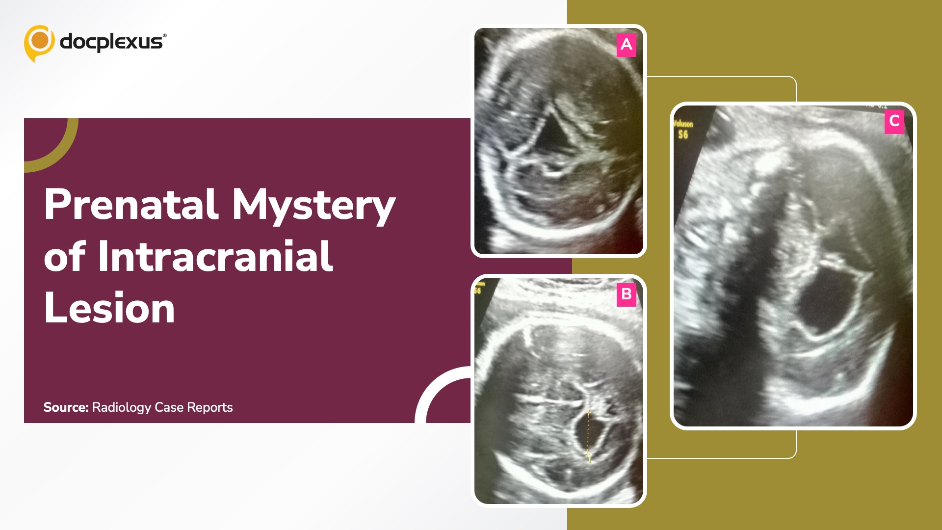
Case presentation A 30-year-old woman in her first pregnancy attended a routine anomaly scan at 24 weeks of gestation. The pregnancy had been uneventful so far, with no history of maternal illness, teratogenic exposure, or hereditary risk factors for congenital malformations. Ultrasound examination A cystic, fluid-filled lesion was identified within the fetal cranial region (Cover Image A).
The lesion was well-defined, anechoic, and lacked vascularity, with a diameter of 15.1 mm (Cover Images B & C). Genetic assessment of amniotic fluid Normal karyotype No chromosomal abnormalities detected Maternal serology Negative for toxoplasmosis and cytomegalovirus Postpartum evaluation of the neonate Cranial ultrasound A well-circumscribed anechoic lesion in the same cranial region Neonatal MRI Nonenhancing, fluid-filled cyst with smooth margins Reference : Radiology Case Reports ##Disclaimer##…



