
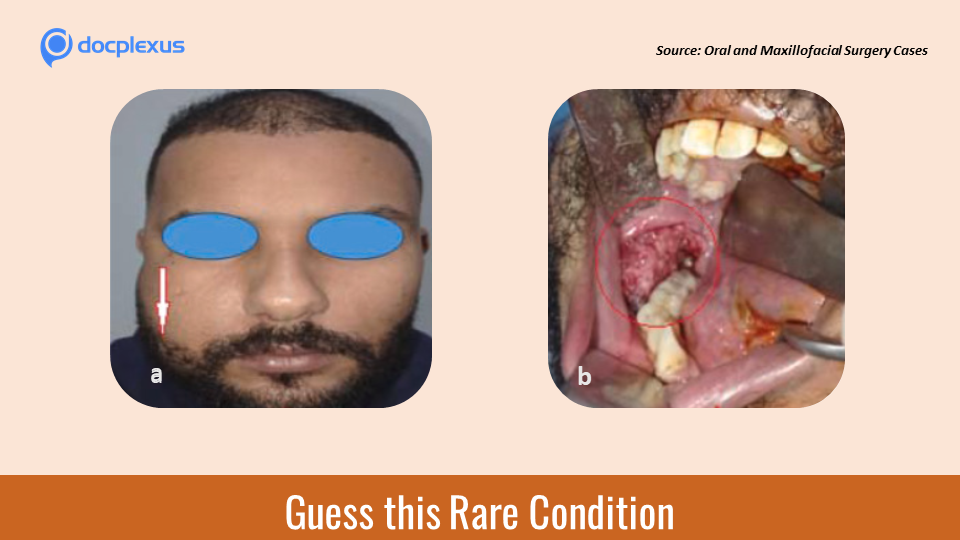
Case presentation A 25-year-old patient reported to the department of maxillofacial surgery for right cheek swelling with no medical history, in particular. The patient complained that the swelling has been gradually increasing in size for the past three months. Clinical examination On palpation, the swelling was firm, fixed, painless, and measured 5 cm x 6 cm (Cover image a). The overlying skin was normal. Endo-buccal examination revealed a raised lesion of the right mandibular gingiva.
Ipsilateral cervical adenopathy measuring 2.5 cm in the longest axis (Cover image b) Radiological investigation The X-ray revealed an osteolytic image showing a mandibular angle extending to the ramus (Figure 1) CT confirmed a tumor in the mandibular axis (Figure 2) Figure 1 . Osteolytic area of right hemi-mandible from 46 to the mandibular ramus, with rhizalysis of 47. (R – Right). Figure 2 . 3D…





