
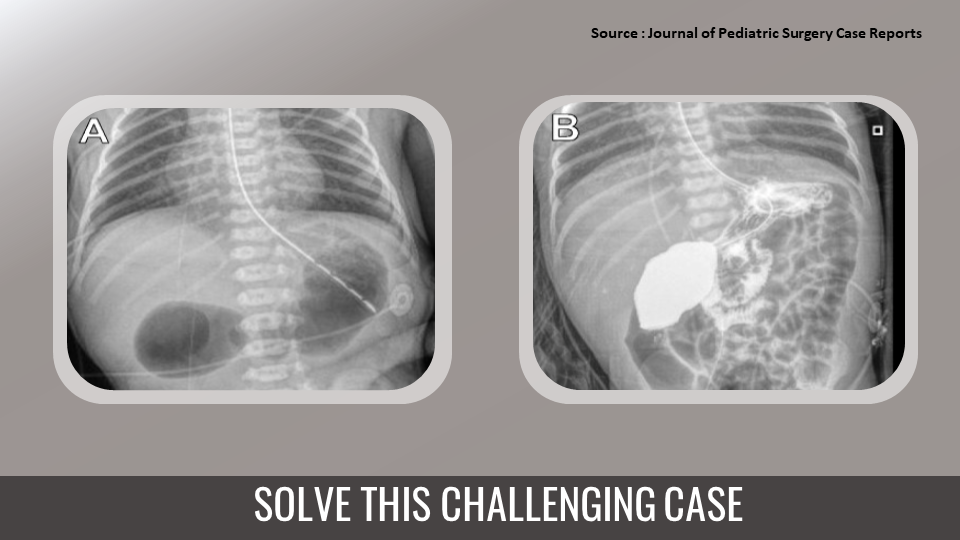
Solve this exciting clinical case specific to your field of interest via Docplexus’ Clinical Case Challenge. Join your fellow doctors to discuss and answer today’s challenge. Case presentation A newborn girl born to a twenty-five-year-old mother at 36 weeks gestation, gravida 3 para 3 was initiated with polyhydramnios and ultrasound suggesting duodenal atresia in the first trimester of prenatal care. The patient had clear emesis, and an orogastric (OG) tube was placed with a return of clear gastric output on delivery.
On day three, post-birth duodenoduodenostomy was performed. Radiological examination revealed Abdominal x-ray with classic “double bubble sign” (Cover image A). Cover Image B showed an upper gastrointestinal contrast study with the passage of contrast through the anastomosis and into the jejunum. Post-operative abdominal x-rays continued showing dilated proximal duodenum…




