
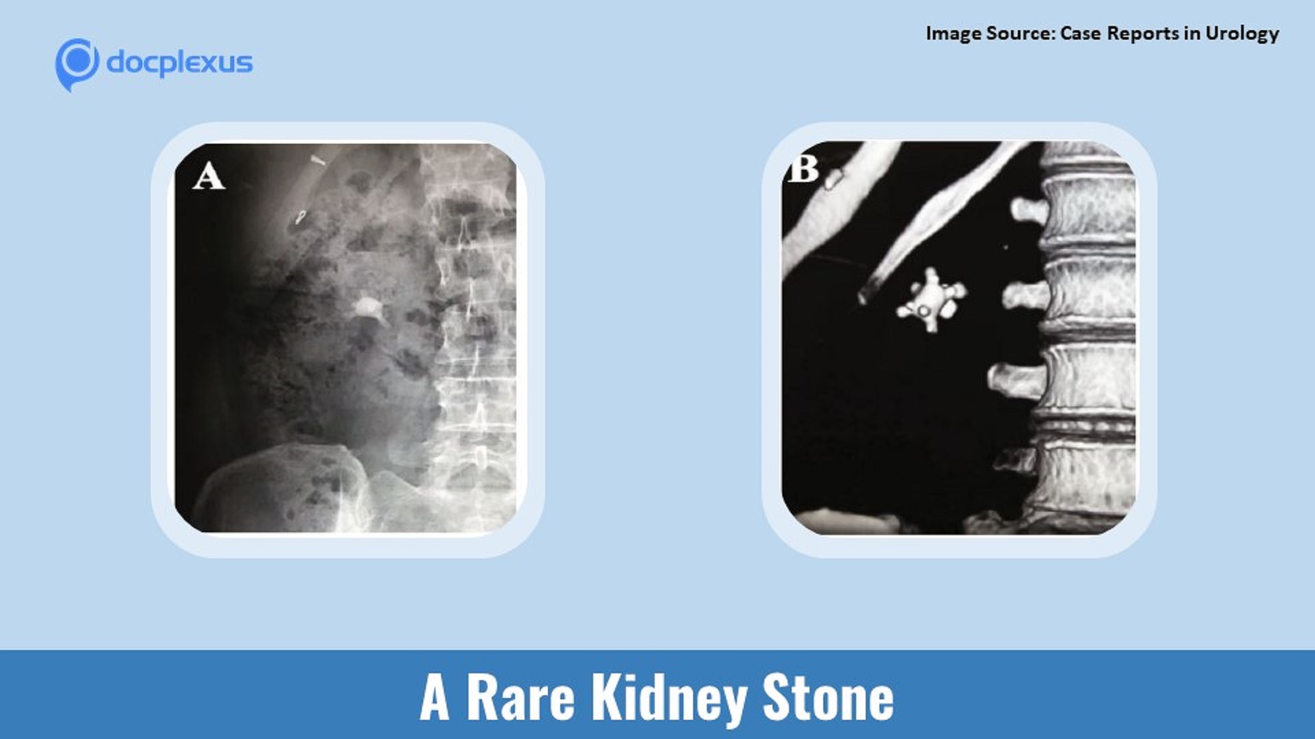
Solve this exciting clinical case specific to your field of interest via Docplexus’ Clinical Case Challenge. Join your fellow doctors to discuss and answer today’s challenge. Case presentation A 63-year-old woman presents with painless, intermittent haematuria for four months. There is no history of flank pain, vomiting, and fever.
She had type II diabetes mellitus for the past 18 years. Her clinical examination is unremarkable. Radiological findings X-ray Kidney Urinary Bladder (KUB): A typical stone in the right kidney (Cover image A) Noncontrast computerized tomography-KUB (NCCT-KUB): Confirmed the presence of a typical stone in the renal pelvis of the right kidney with mild hydronephrosis and no proximal hydroureter. Stone size was 2.4 cm x 2.3 cm, and stone density was 1355 Hounsfield units (Cover image B) Diuretic renography: Ruled out significant obstruction with 45% right kidney…





