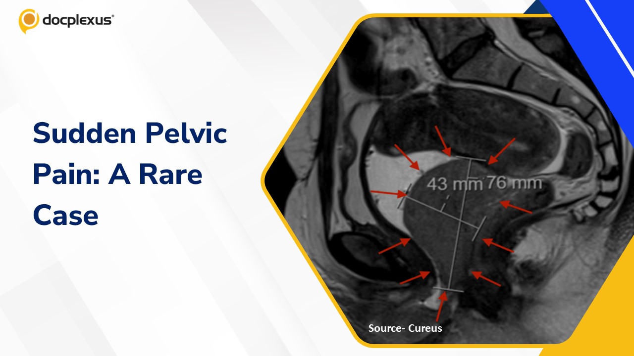
Case Presentation A 41-year-old woman with a history of two cesarean deliveries and no known comorbidities, presented with progressive pelvic pain and dyspareunia persisting for three months Physical examination On pelvic examination, a firm, fixed mass was palpable in the posterior vaginal wall. There were no associated symptoms such as abnormal bleeding or discharge Diagnostic Assessment Laboratory Investigations Hemoglobin: 13.7 g/dL White blood cell count: 10.08 × 10⁹/L Platelet count: 325 × 10⁹/L All initial hematological parameters were within normal limits Imaging Studies Transvaginal Ultrasonography: Revealed a solid lesion measuring 7 × 5 cm in the posterior vaginal wall (Cover Image) Pelvic MRI: Demonstrated a 7.5 × 4.3 cm solid mass in the rectovaginal space without rectal invasion The lesion caused anterior compression of the bladder and posterior displacement of the rectum,…
A Rare Twist: Sudden-Onset Pelvic Pain in a Young Woman

Case Challenge
Medical Writer