
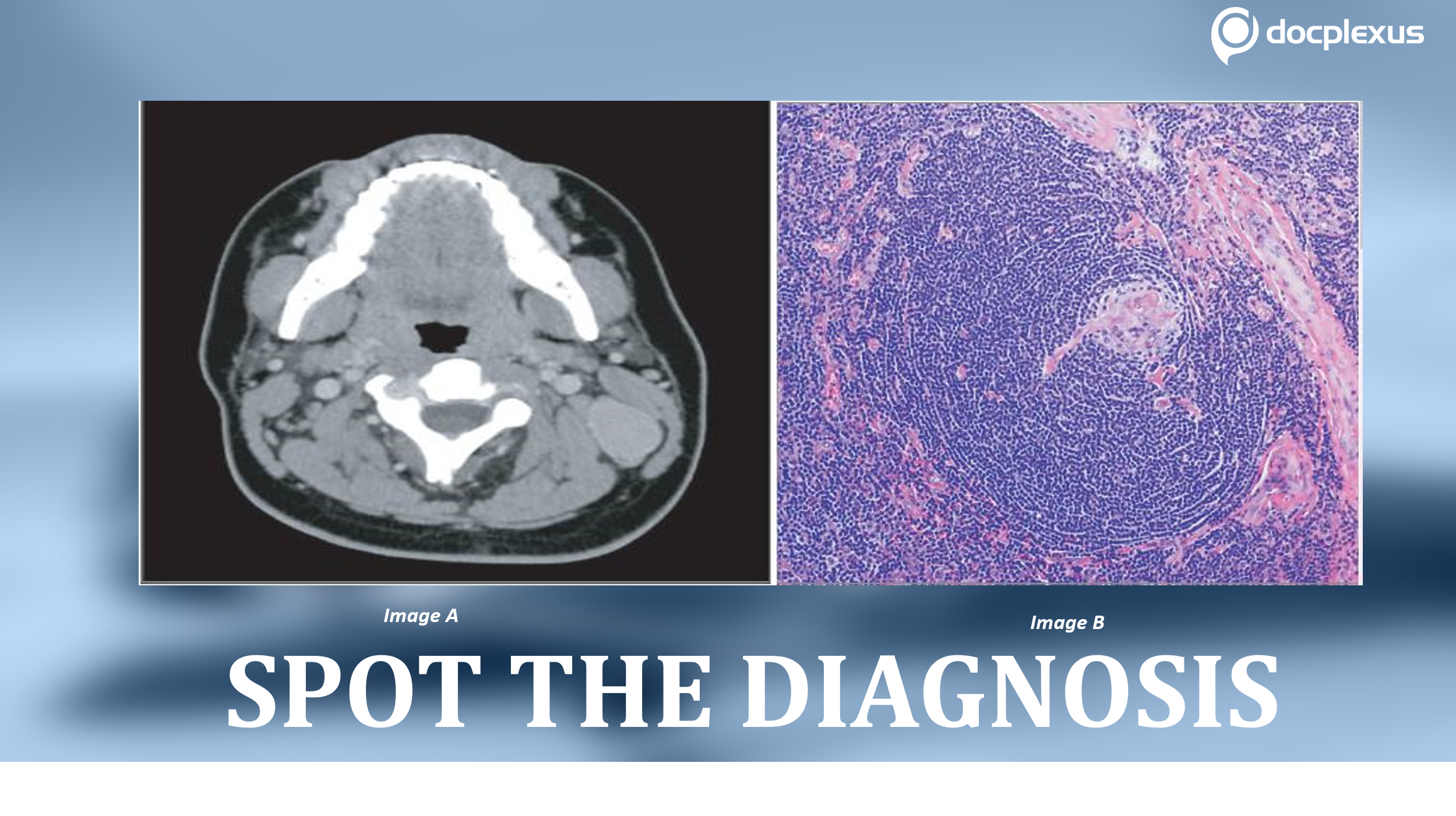
A 17-year-old male was presented to a specialty clinic for the consultation of a left-sided neck mass persistent for the last six months. Earlier, the patient had presented to the primary care center, and the ultrasonographic image revealed a 4.0 x 1.5 x 2.8 cm lymph node level 2 on the left side. The patient received antibiotic treatment, but it did not improve the condition. Subsequently, he developed a fever and sore throat.
During his present consultation, an axial CT scan of the neck was performed (Image A) that revealed an isolated, homogenously enlarging left-neck lymph node of level 2/5A. The patient reported no relevant clinical or surgical history. Biochemical analysis White blood cells: 11310 cells/µL C-reactive protein: 56.2 mg/L The tests for influenza A and B, HIV, cytomegalovirus, and Epstein-Barr virus showed negative results. Ultrasonography-guided needle biopsy,…


