
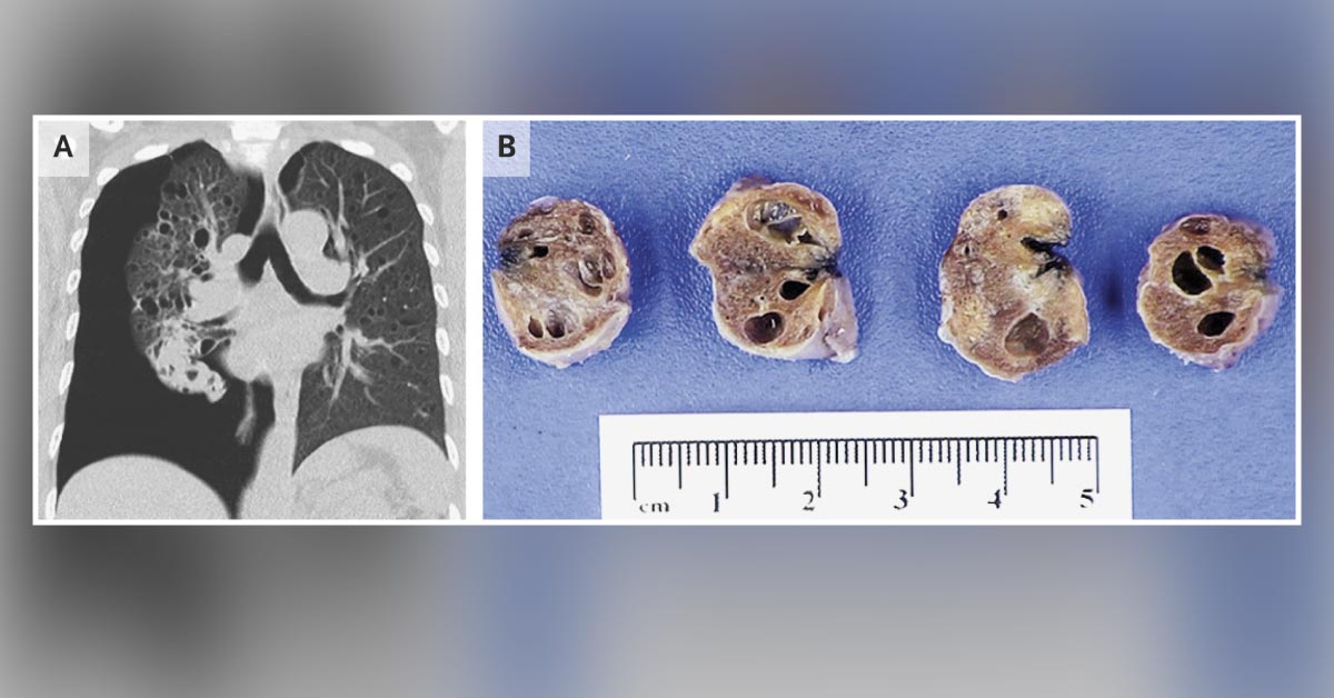
Solve exciting clinical cases specific to your field of interest via Docplexus clinical case challenge. In today’s ‘Pulmonary Case Challenge’ join your fellow doctors to discuss and answer an interesting quiz. A 44-year-old woman presented to the emergency department with acute chest pain after several months of progressive dyspnea. Her oxygen saturation was 92% while she was breathing ambient air, and the physical examination was notable for diminished breath sounds on the right side.
Computed tomography of the chest, reconstructed in the coronal plane at the level of the tracheal carina, revealed a large pneumothorax and diffuse, intraparenchymal pulmonary cysts (Image A). The patient underwent mechanical pleurodesis, and a wedge biopsy was performed. The biopsy specimens contained numerous thin-walled cysts (Image B). Source: The New England Journal of Medicine





